Time is brain
CBV, CBF, MTT, TTP and Tmax perfusion maps are computed in seconds: the physician sees the infarct core and penumbra and decides on thrombolysis or thrombectomy faster.
The Kometa neurology solution brings CT brain perfusion, multi-sequence MRI, head and neck angiography and PET/CT into one workspace connected to the archive, the RIS and the regional teleradiology network.
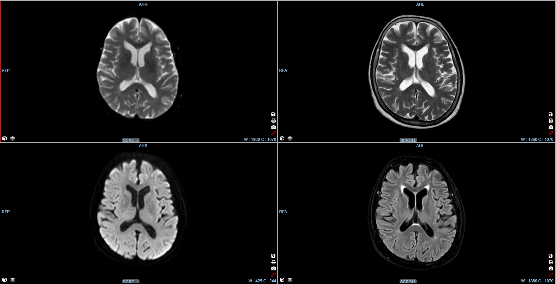
CBV, CBF, MTT, TTP and Tmax perfusion maps are computed in seconds: the physician sees the infarct core and penumbra and decides on thrombolysis or thrombectomy faster.
DWI, FLAIR, T1, T2 and post-contrast series open synchronised: scrolling, window level and zoom are linked across viewports, and the prior study is one click away.
A study from a district hospital opens for the neuroradiologist of the regional centre in a browser. The Kometa regional system runs in 29 Russian regions.
Layouts for stroke, multiple sclerosis, epilepsy and CNS tumours; measurements, annotations and key images are saved into the report.
Neuroradiology is one of the core areas of Kometa Workstation MRI and CT. The perfusion and multi-sequence MRI tools were developed together with radiologists working in stroke centres and neurology departments.
The clinical product-integration practice, the R&D department and technical support take part in every deployment: they configure protocols for the stroke centre, connect the equipment and train staff on site.

Methodologists and consulting physicians define the requirements for perfusion and MRI protocols and help build the stroke pathway between the primary stroke unit and the regional centre.
Our own development team: perfusion maps, series synchronisation and 3D / MPR tools evolve on request from clinics.
Kometa 3Di PACS regional archives connect hospitals and stroke centres: teleradiology and expert review are available from any point of the network.
Each stage covers its own part of the journey — from the first CT in the emergency room to expert review and the report in the RIS.
Non-contrast CT, CT angiography and CT perfusion for suspected stroke arrive in the archive right after scanning.
Learn moreSeries and the patient's prior studies are available in Kometa PACS — in the hospital, the cloud or the regional archive.
Learn moreKometa Workstation: CBV / CBF / MTT / TTP / Tmax maps, MRI series synchronisation, vessel MIP and 3D, measurements.
Learn moreThe neuroradiologist of the regional stroke centre opens the study in WEB-Viewer and confirms the treatment strategy.
Learn moreThe report in Kometa RIS, transfer to the HIS, REMI and the regional network; follow-up compared against the archive.
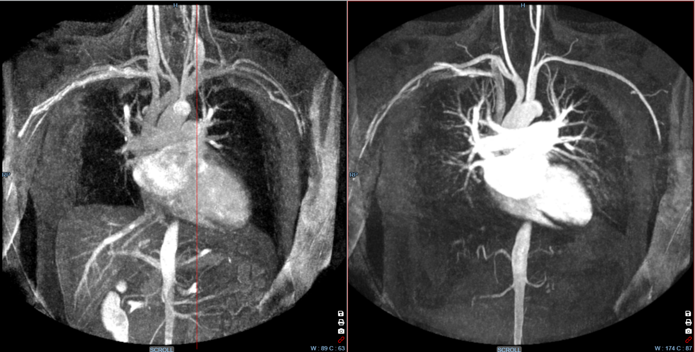
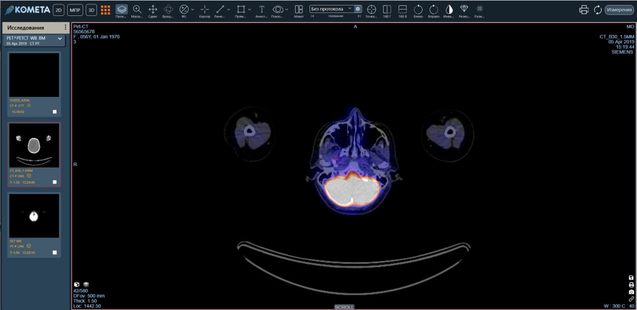
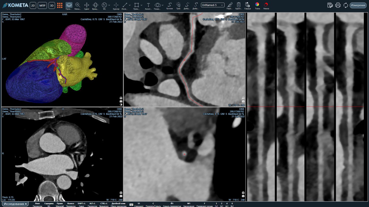
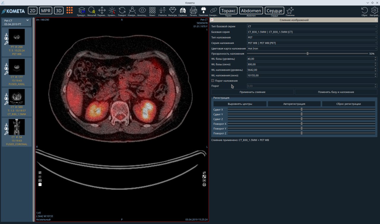
Learn moreThe neuroradiology modules of Kometa Workstation CT and MRI, shown on real screens of the system.
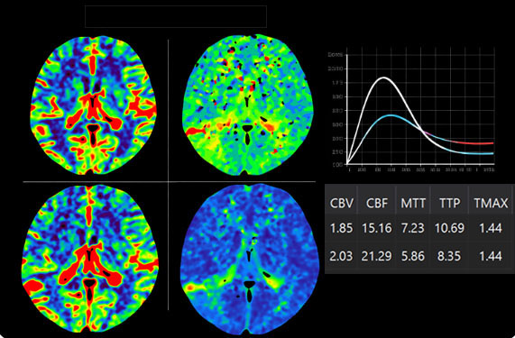
CBV, CBF, MTT, TTP and Tmax perfusion maps computed from dynamic CT, with arterial and venous curves and a table of values per region of interest.
Professional MRI diagnostic workstation optimized for neurological, musculoskeletal, and oncological imaging.
Learn more →Professional CT diagnostic workstation with advanced multi-planar visualization and measurement tools.
Learn more →
Browser-based DICOM viewer with WebGL acceleration — high-performance diagnostics with no software installation.
Learn more →
Centralized regional PACS connecting multiple medical facilities into a single unified network.
Learn more →
Radiology Information System for managing patient schedules, referrals, and structured diagnostic reports.
Learn more →Kometa products are built on the DICOM standard and registered for medical use in Russia. This guarantees interoperability with modalities, PACS, RIS and the hospital and regional information systems.
Kometa 3Di PACS — registration certificate No. RZN 2017/6377.
Entry No. 2300 of 15 December 2016 in the unified registry of Russian software.
Certified quality management system.
Integration with any modality, HIS, RIS and AI services; DICOM Worklist support.
Maps are built on the workstation within seconds after the arterial and venous points are selected; no separate computation servers are needed.
We will show perfusion and MRI tools on your own studies and propose a configuration — from the emergency room to regional teleradiology.
Software product intended for use by medical professionals.