

Kometa Workstation MRI
Kometa Workstation MRI is a multifunctional workstation that works with any digital medical equipment: CT, MRI, ultrasound, and other devices. The solution allows for efficient organization of diagnostic image viewing and analysis, and accelerates clinical decision-making.
The intuitive interface and rich toolset ensure rapid deployment in radiology departments, increasing team productivity and study quality.
Instructions for use are available for download in the Documents section.
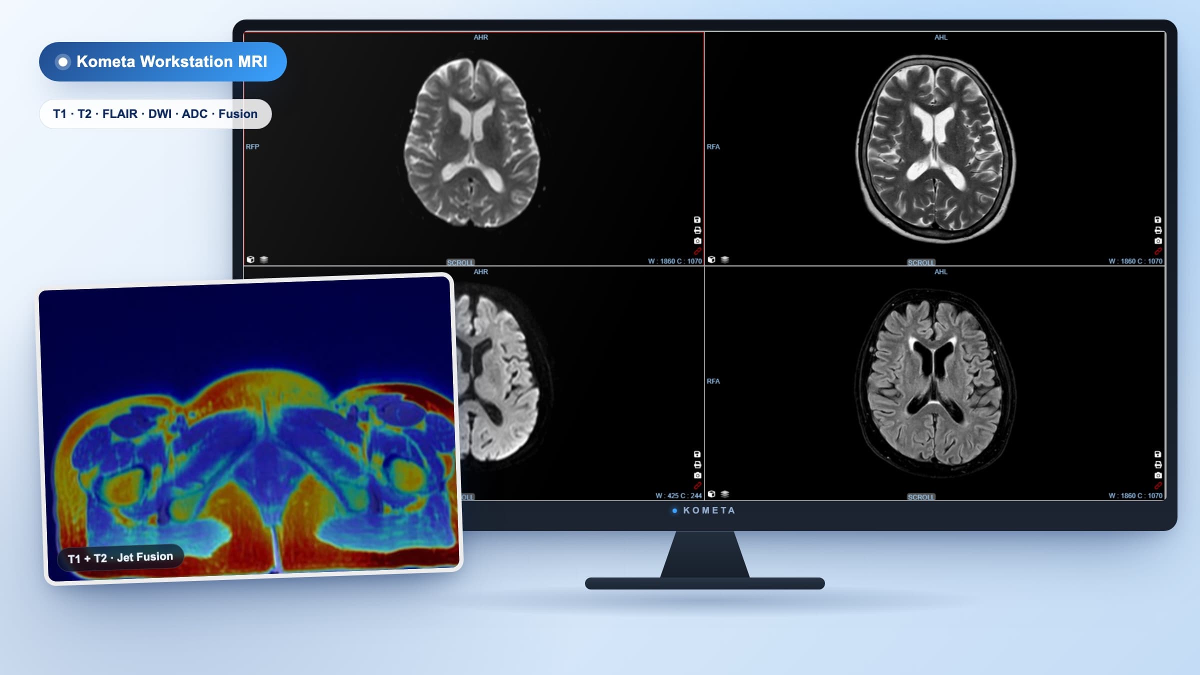
Kometa Workstation MRI capabilities
Kometa Workstation MRI combines visualization, processing, and analysis, helping physicians get results faster and improve diagnostic quality. Below are the key advantages.
Local storage and processing
Instant access to saved data with no limits on the number of views and actions.
Fast study search
Convenient filters and results by key patient and protocol parameters.
Specialized modules
Additional applications for study analysis across each diagnostic profile.
Visualization protocols
Choose base templates or create your own protocols with saved presets.
Maximum visualization tools
MPR, MIP, and 3D reconstructions in any projection at one workstation.
Clinical applications
Oncology, cardiology, neurology, dentistry, and other fields in a unified interface.
Precise measurements
Distances, angles, tissue densities, organ sizes, and lesions of any shape.
Markers and notes
Add markers and save processing results for colleagues and follow-up studies.
Layer-by-layer tissue removal
Structure segmentation algorithms help focus on the target analysis zone.
Cine series playback
View dynamic series and control playback directly in the work window.
Multi-monitor support
Connect displays of different sizes and resolutions with individual settings.
Flexible interface
Customize window layout, color palette, language, and hotkeys to your preferences.
Integration with Kometa Classic PACS
Instant data exchange with the archive and other Kometa ecosystem solutions.
RECIST and PERCIST analysis
Objective assessment of lesions and treatment dynamics by international standards.
Data security
Authorization by username and password, access rights management, and user action control.
Who is it recommended for?
All healthcare facilities working with medical images. Kometa Workstation MRI deploys as part of a comprehensive ecosystem or in fully autonomous mode. In both cases, the workstation handles visualization, analysis, and storage of study results. Thanks to deep integration capabilities, results are easily exported as printed reports, images, or to the archive.
Kometa Workstation MRI complements every Kometa Storage Edition product and deploys autonomously in any radiology office. The workstation covers the complete physician workflow: from receiving studies to processing, comparison, and result preparation.
Ready to see Kometa Workstation MRI in action?
Submit a demonstration request, and we'll configure the demo according to your department's tasks. A specialist will discuss implementation, integration, and support scenarios.
Illustrations and screenshots
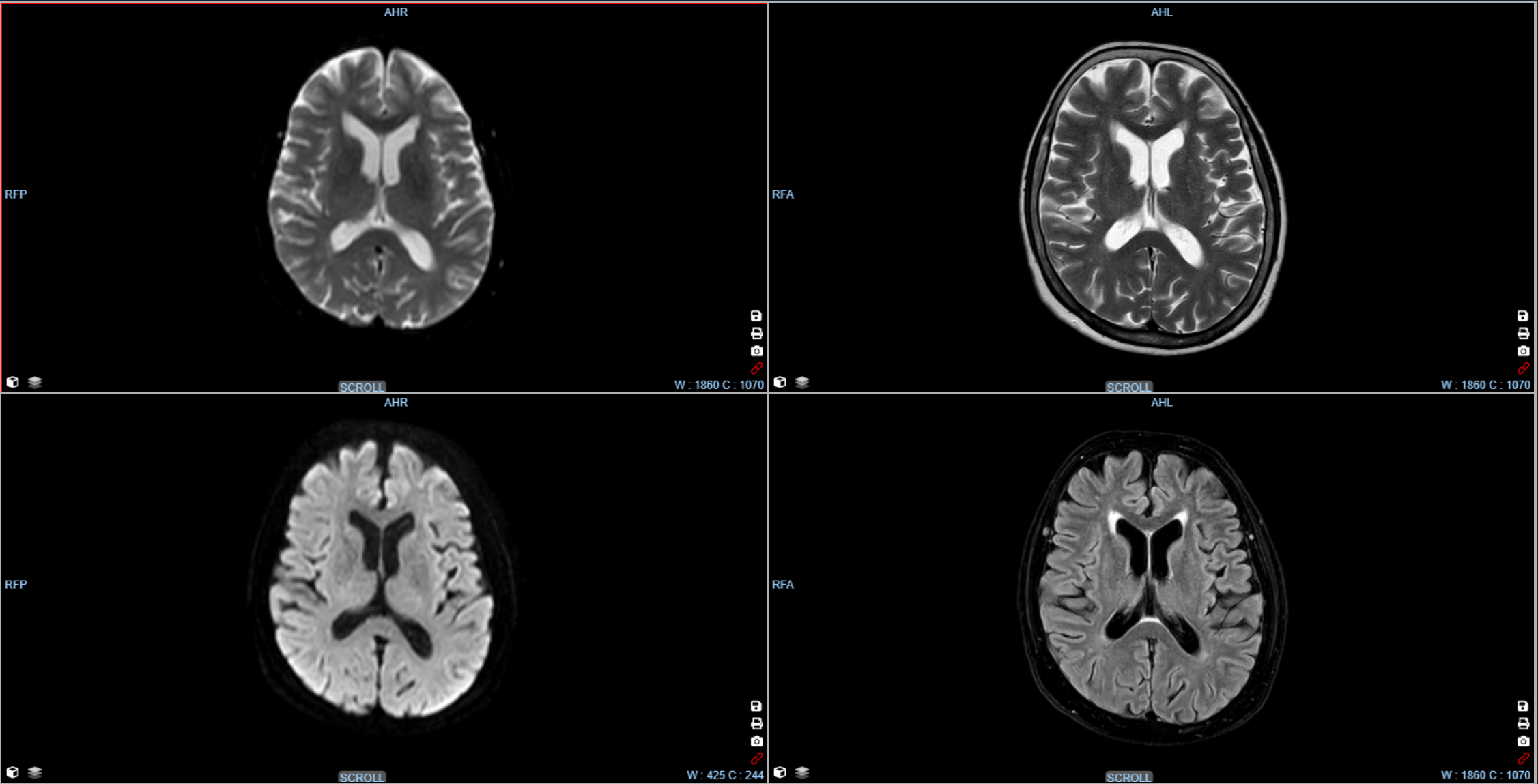
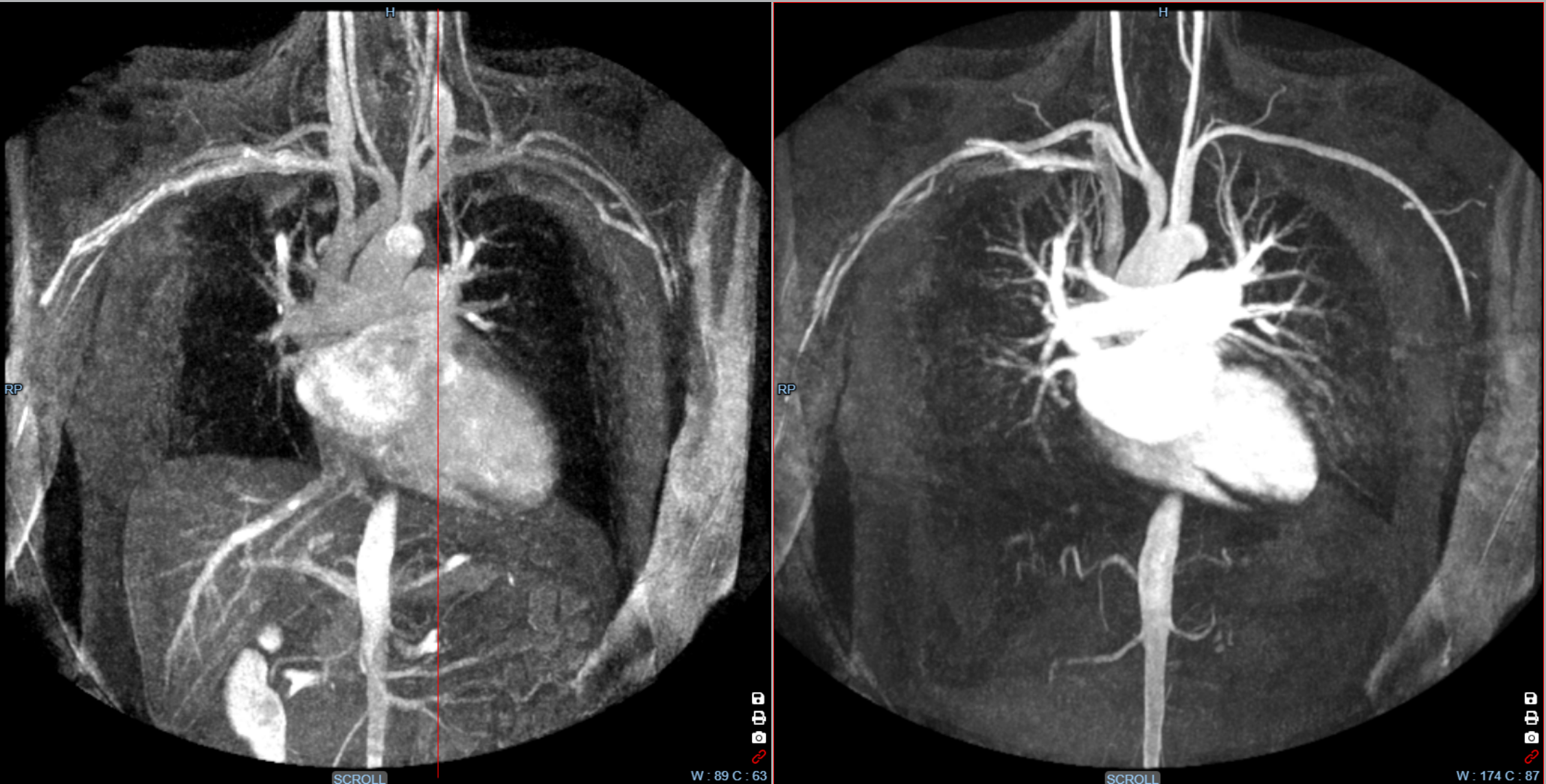
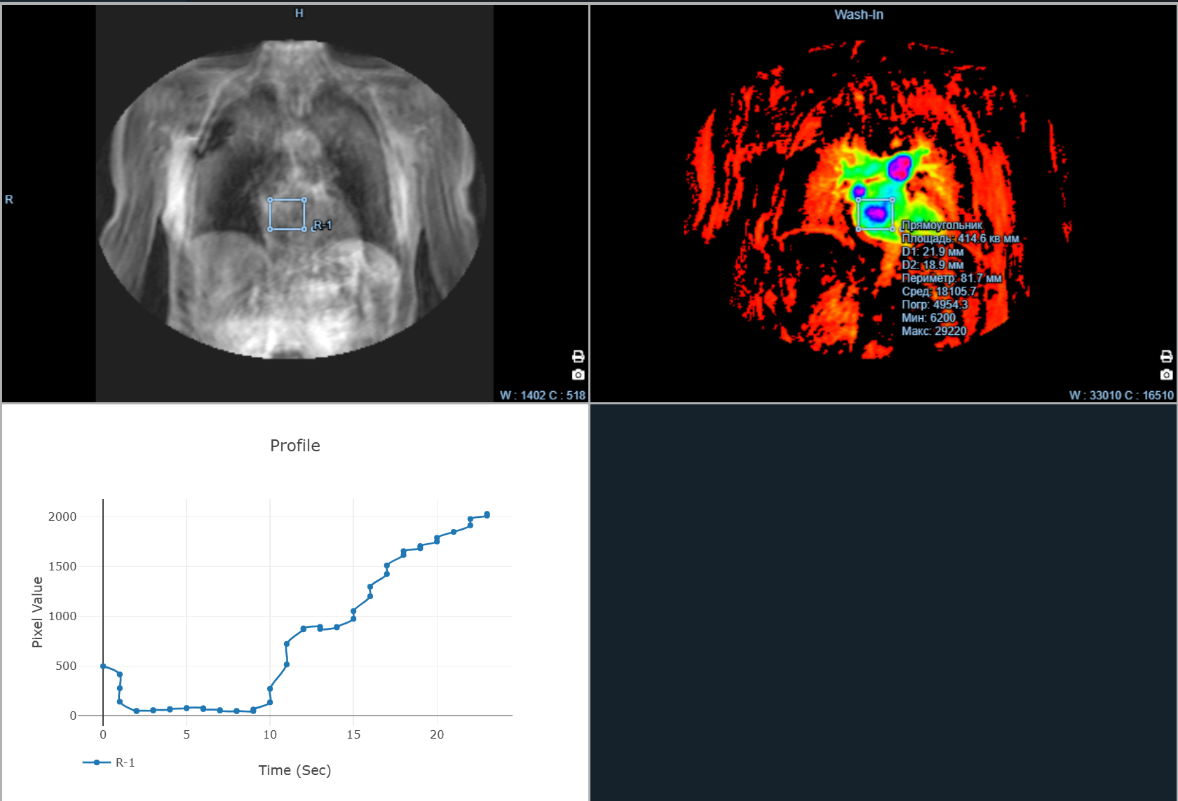
Product modules
Expand the workstation with specialized modules. Combine them for specific clinical tasks and create a unique capability stack for the radiology team.
Modules
Pair 1 (T1 + T2 / Jet)
Fusion of T1 and T2 series in the axial plane with the Jet colormap.
Gallery
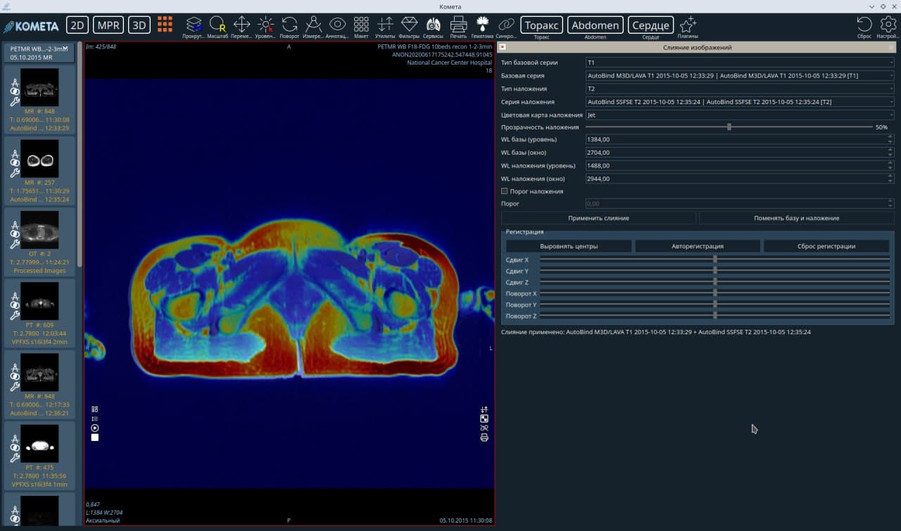
Pair 1 (T1 + T2 / Jet)
Click to enlarge
Pair 1 (T1 + T2 / Jet)
Fusion of T1 and T2 series in the axial plane with the Jet colormap.
The image shows fusion of two MRI series in the axial plane: the base layer is T1 (AutoBind M3D/LAVA T1) in grayscale, and the overlay is T2 (AutoBind SSFSE T2) with the Jet colormap at 50% opacity. Jet maps T2 intensity from blue through green and yellow to red, enabling direct anatomical correlation of both sequences in one view.
Creating a digital environment for diagnostic physicians
Kometa Workstation MRI is a reliable tool for those who work with diagnostic images every day. The workstation helps you focus on what matters most — the patient, not routine operations. Contact us to discuss implementation and find the optimal configuration.
Software product intended for use by medical professionals.
Video reviews
Watch overviews of Kometa products and modules
3D mode, segmentation — KOMETA 4.1.21 Viewer
3D mode, segmentation — KOMETA 4.1.21 Viewer
WEB Viewer 4.1.21 interface overview
2D mode — KOMETA 4.1.21 Viewer
MPR mode, comparison package — KOMETA 4.1.21 Viewer
VESSELS package — KOMETA 4.1.21 Viewer
Endo package — KOMETA 4.1.21 Viewer
Viewer Kometa Pathology
Cardiac package — KOMETA 4.1.21 Viewer
Calcium package — KOMETA 4.1.21 Viewer
PET-CT package — KOMETA 4.1.21 Viewer
Lungs package — KOMETA 4.1.21 Viewer
Brain perfusion package — KOMETA 4.1.21 Viewer
Dental package — KOMETA 4.1.21 Viewer
Radiation Therapy package — KOMETA 4.1.21 Viewer
Mammography — KOMETA 4.1.21 Viewer
Web Patient Browser overview